Estimated read time: 4-5 minutes
This archived news story is available only for your personal, non-commercial use. Information in the story may be outdated or superseded by additional information. Reading or replaying the story in its archived form does not constitute a republication of the story.
ST. GEORGE — Brain surgery is one of the most serious procedures someone might undergo, which is why it's so important doctors have every resource available to them. One Utah man credits his life to a technology that's new to St. George.
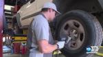
Keith Barney is a 28-year-old mechanic in Cedar City.
"I love figuring out broken things and taking them apart and putting them back together,” he explained.
But after hitting his head on a ball hitch while working under a truck, Keith discovered a problem he couldn't fix on his own.
"I've hit my head thousands of times before but never this hard,” he said.
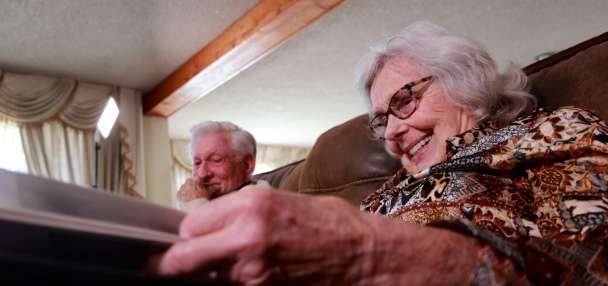
With a little encouragement from his wife, Kimberly Barney, he reluctantly went to the emergency room for an MRI only to learn he had a brain tumor.
"It was literally a golf ball!” Keith Barney described. “And we're like, ‘Oh man, this is pretty serious.’”
Kimberly Barney was prepared for the worst. "I was pretty much terrified,” she said.
Keith Barney went in for 16 hours of surgery in a specialized operating room, called a dual-purpose intraoperative MRI suite. The setup allows the doctor to take an MRI during the middle of brain surgery to measure progress and eliminating the need for multiple surgeries.
Intermountain Healthcare's Dr. Berkeley Bate, a neurosurgeon at Dixie Regional Medical Center, said real-time imaging throughout surgery helps him identify and remove the tumor more accurately.
“These tumors, especially low-grade gliomas, they can look a lot like normal brain (tissue),” he said.
In the past, Bate had to delay taking an MRI until after surgery, occasionally realizing parts of the tumor were still there.
More Your Life Your Health:

St. George woman loses 105 pounds after having bariatric weight-loss surgery
It's easy to set the same New Year’s goals year after year and not see much progress. After dieting and exercising without seeing results, one St. George woman decided it was time to make a bigger change.“Sometimes there are chunks left, or there are pieces hiding and you don’t see it, but you can see it in the MRI. You take them back to surgery that next day or two days later, and open all back up again,” he said.
Bate said the new setup in the suite is convenient and nice for the patient, too, since they don’t have to go back to surgery.
“Whereas with this, it's the same thing, but they're asleep. So, you just slide them off this table onto the MRI table, wheel them in and get an MRI,” he explained. “And then I just have the radiologist come up and look at the pictures with me … and then I make a judgment call. ‘OK, am I done? Or do I need to keep going and take some more out?’”
Bate said this new system also helps doctors be extra careful around structures that control motor and speech functions.
“You want to be aggressive and get everything out without being so aggressive that you cause a neurologic problem,” he said.
Bate said the neuronavigation system on screens in the operating room allows him to match the patient’s brain and real space from the picture.
Kimberly Barney was worried about her husband before he went into surgery. "I wasn't sure if he was going to be OK, or if he would even remember who I was,” she said.

Fortunately, Keith Barney's surgery was successful.
"I was happy with them taking their time to make sure they got everything out without having to go back in and cut his head open a second or third time,” Kimberly Barney said.
“If we can get all that tumor out safely without causing a problem for him, then he's got a lot better chance of living and enjoying his life,” Bate explained. “Because if you cause a neurologic deficit, he’s got to live with that for a long time.”
Keith Barney said the doctor told him the tumor could become cancerous if it wasn’t taken care of when they removed it. “I was a few months away from having brain cancer,” he said.
After surgery, Keith Barney said he was home in just six days. “Which is pretty freakin' amazing," he said.
He said he went to a few sessions of occupational, physical and speech therapy but had a relatively easy recovery. Today he is back to work and is able to drive again.
"I wake up every morning and thank God I don't have a brain tumor,” Keith Barney said.
His wife is equally thankful. “We tell each other every day just how grateful we are that he's OK and he's still here,” Kimberly Barney said.
“It's a big deal to have something like this in a town like St. George,” Bate said. “I think it really helps us provide world-class care.”
Primary Children's Hospital also has plans to build an intraoperative MRI suite in the future. It is currently finalizing construction design.