Estimated read time: 2-3 minutes
This archived news story is available only for your personal, non-commercial use. Information in the story may be outdated or superseded by additional information. Reading or replaying the story in its archived form does not constitute a republication of the story.
Ed Yeates ReportingPhysicians have unwrapped some new 3D technology to search out subtle problems of the heart that often go undetected. The harmless non-invasive tool is revealing some things that, up until now, required driving an invasive catheter into the heart.
Though 19-year old Jeff Addy is an avid hiker, when he climbed to the top of King's Peak on Saturday, he started feeling weak, something he had never experienced before.
Jeff Addy: "Real hard time breathing, getting tired easily – headache, kind of chills, cough."
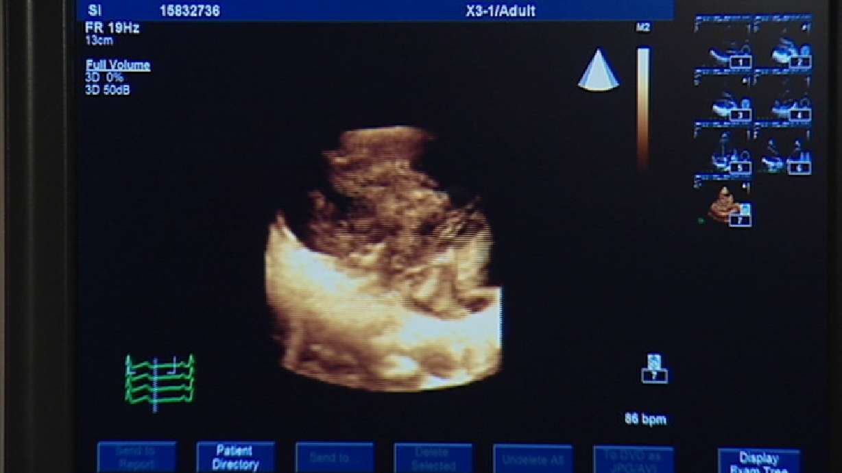
The symptoms are typical of altitude sickness, but just to make sure, doctors at the University of Utah Hospital put his heart under the non-invasive probe of a new 3D ultrasound.
In 2-D, this is about all you can see of the left ventricle of the heart. But in three dimension, all the detail comes out, including what is affectionately called the fish lip movement of the valve. Jeff certainly could see the difference.
Jeff Addy: "Well, it's just amazing. You can see the texture in so much more detail. Kind of get more of an idea what it really looks like."
From the bottom looking up, to the top looking down, and rotating, the 3-D moves around and through and into areas sometimes hiding subtle changes or damage. And it's all in REAL time.
Using the same technology, researchers can also set up parameters to literally map all the intricate movements of the heart, even the kind of electrical movements requiring positive and negative charges to and from the heart muscle - which sometimes get out of whack - causing an irregular or erratic heart rhythm.
Kevin Whitehead, M.D., University of Utah Cardiologist: "What an advantage to see all of the walls of the heart beating at the same time in one view and know where the areas are that need to be sped up to catch up with the rest of the heart and beat in a more coordinated fashion."
The pressure of fluid movements to and from the heart are shown in colors. It also shows valve performance - the flexing of the heart muscle - even hypertension in the lungs.
Jeff may or may not have any of these subtle things going on, but if he does, this 3-D ultrasound will seek it out.


